Activity
Mon
Wed
Fri
Sun
Dec
Jan
Feb
Mar
Apr
May
Jun
Jul
Aug
Sep
Oct
What is this?
Less
More
Memberships
MSK School (Free)
Public • 822 • Free
3 contributions to MSK School (Free)
TENNIS LEG CASE
Hi everyone , i m new in this comunity , big fan of Agten Radiology on Youtube, here a case from my Linkedin profile . Tennis leg case are always very challenged for me , so if you have cases to share i ll be grateful. Coronal and Axial T2-weighted fat-suppressed images shows a hyperintense fluid collection between the left medial head of the gastrocnemius and soleus ( purple arrows) There is partial tear of the distal medial head of gastrocnemius with some retraction( red arrows) which extends to a few fibers of the lateral head ( blue arrows) Plataris tendon was intact.
1
3
New comment Jul '23

1 like • Jul '23
@Matvei Shiltsin tennis leg was use for plantaris tendon lesion that is asocciated with fluid in the fascia between soleus and medial gastrocnemius, but then was discovered that most of times there was lesion of medial gastrocnemius or soleus muscle , so now tennis leg is use for lesion of the calf muscles , sometimes just one or combinated ... more info in https://radsource.us/tennis-leg-plantaris-tendon-rupture/ AND https://radsource.us/not-plantaris-keys-better-diagnosis-calf-strain-injuries/
Shoulder case
38 year old female with shoulder pain and swelling of 1 week. patient had difficulty moving the joint.
4
13
New comment Jul '23

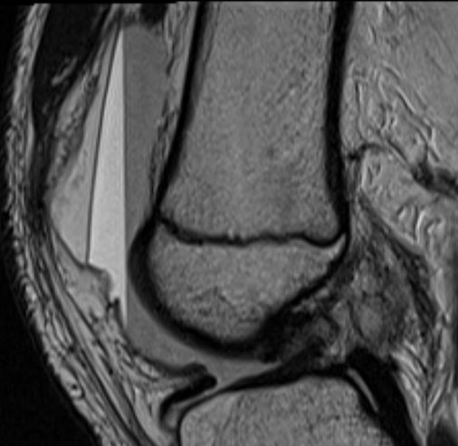
Lipohemarthrosis and gravity
This image shows a lipohemarthrosis after patella dislocation. 3 layers of fluid. what happended to gravity. What do you think? Answer with an explanation, why you think this happens, can be funny and not scientifically validated. GIF answers allowed :)
Complete action
6
16
New comment Jul '23

1-3 of 3
@claribel-balsano-6714
MD, Radiodiagnosis especialist from Buenos Aires Argentina
Active 481d ago
Joined Jul 4, 2023
powered by


